#mitochondria
#MMDATA
#GUIDE
Mitochondria Guide Marking During All Time Activity
Cut-off – “Just the central image of the flipbook please – the other images will become the central slice in other flipbooks so all images will be drawn on eventually. Thanks for your help!” LucyEM
SHADOW – “shadows would appear like the dark edge of the mitochondria. So you need to draw around the shadows if them if you can see them, but only if you can tell they’re part of the mitochondria in an adjacent slide.”, mattrussell
single or two separate – This actually appears to be a site of mitochondrial fission or fusion; the thin tubes you can see touching the point between the mitochondrial lobes are parts of the endoplasmic reticulum, which has been shown to surround and possibly drive fission/fusion at these sites (see papers by the Voeltz lab at CU Boulder), mattrussell
When are regions enclosed in membrane not mitochondria? circle are lipid droplets and not mitochondri, the red marking are mitochondria
You can see similar structures with a similar ‘halo’ of stain in figure 1 here and figure S1 at this link here; opens a PDF. “mattrussell”
Where to find the mitochondria guide of marking?
Are there any here? No, this subject hasn’t any mitochondria. “Remember that some images may not have any mitochondria in them. It looks like this is just the edge of the cell.”, mattrussell
Is there 10? No, much more:
How about MISTAKES?

Subject 50730439
Red marks are including the mitochondria’s shadow due to 12345 (typical mitochondria motion, fusion and change and you can to see the mitochondria membranes, ‘sometimes you can’t to see membranes due to 3D to 2D view, when they are on a other other side (axes Z like graph xyz) and in this case you needs to do another research (find them in the other slides 12345 or on the another similiar near subjects). This research is important, because some aren’t mitochondria, but LIPIDs etc. – mitochondria often change shape rapidly and move about in the cell almost constantly and sometimes is on the same place without shaping etc.)


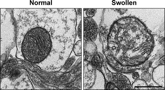
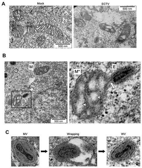
hello everyone, I did some verification about the mitochondria degradation, because a lot of subjects looks similarly like these:
I saw before something like swalling, virion, etc.
A, B. Representative mitochondrial swelling


Altered mitochondrial morphology in cumulus cells of diabetic mice
infection alters, Punctate mitochondria co-localized with progeny virions interact

